Brain Ventricles Neurology

Brain Plumbing
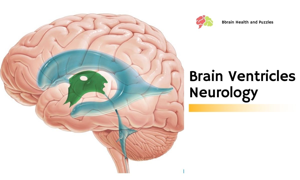
In terms of brain ventricles, neurology has come a long way in describing their purpose, knowing when they are normal and when they are abnormal. There are several different brain ventricles neurology has named based on their position and brain anatomy. There are two lateral ventricles, one on the right and one on the left. These brain ventricles are connected to the third ventricle which is centrally located.
There are no first or second ventricles because of the existence of the lateral ventricles. Since they are symmetric, a numbering system was not used for the lateral ventricles. The third ventricle is connected to the fourth ventricle by the cerebral aqueduct.
Eventually, these ventricles connect with the spinal cord. This system of brain ventricles allows cerebrospinal fluid to flow through the brain and spinal cord like water flowing through basins and pipes. Instead of a pump to push the fluid, the cerebrospinal fluid is constantly produced by cells in the brain ventricles called ependymal cells. Because the cerebrospinal fluid is constantly made and released into the brain ventricles, it pushes the fluid throughout the system.
A Cushion of Liquid
The brain is unique in that it rests on a cushion of liquid. It is held in place by something that is reminiscent of a hammock or sling and is surrounded by cerebrospinal fluid. Strangely, the brain sloshes around ever so slightly in the skull.
If you have an injury to one side of your head the brain will move with great force and strike the opposite side of the skull. In neurology, this is known as a contrecoup (pronounced kon-tra-coo) injury. Not only will you have damage to the brain directly from the blow to the head, but also on the surface of the brain on the opposite side.
This Is (Where You) Spinal Tap
Sometimes brain tumors can compress on the brain ventricles, especially the very small cerebral aqueduct. This causes a backup of cerebrospinal fluid in the brain which leads to terrible headaches among other symptoms. Surgery is usually required, not only to remove the brain tumor but also to relieve the built-up pressure from the cerebrospinal fluid.
When you have a spinal tap, the doctor is taking a sample of cerebrospinal fluid to test. It is taken from the lower back so that the needle does not injure the spine but can reach the cerebrospinal fluid that is produced high up inside the brain ventricles. By taking a sample of this fluid, doctors can determine if the brain is healthy or sick.

Physicians who specialize in neurology are experts in performing these procedures and analyzing the results. Some illnesses that can be diagnosed with spinal tap (also known as a lumbar puncture) are meningitis, encephalitis, and normal pressure hydrocephalus. Not surprisingly, the field of neurology studies and treats these illnesses. Moreover, without brain ventricles, neurology would have to develop a new way of diagnosing these illnesses.
By measuring brain ventricles, neurology has been able to uncover anatomical clues about dementia. Alzheimer’s disease is the most common type of dementia and it results from nerve cells dying off over time, usually later in life. As these nerve cells die, the brain ventricles grow to take their place.
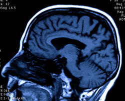
This effect can be seen on CT and MRI scans of the brain. By looking for subtle increases in the size of brain ventricles neurology is making the diagnosis of dementia earlier and allowing treatment to begin faster. Earlier treatment leads to better outcomes.


