Thermal Brain Imaging – A Harmless Peek Inside The Brain

Thermal imaging is an exciting new way for physicians to take a look at the activity inside your brain. It’s completely harmless but is still in the early stages of its development. It has been used with success in certain circumstances but has shown limitations.
The good news is, that its use is being studied and refined all the time so if and when you need your brain scanned look forward to this non-invasive technique.
Hot and Cold
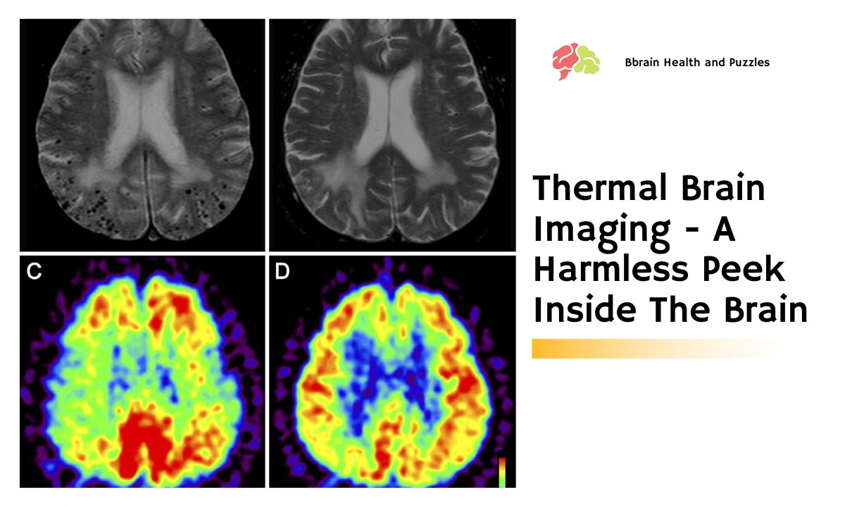
 Thermal imaging is a method of determining how much heat is given off by individual areas of the brain. These hot spots and cold spots show up on a computerized image of the brain as red or blue areas. These areas are displayed by means of infrared photography. It’s the same sort of concept that’s responsible for night vision goggles, those kinds often used by hunters, or the military.
Just a few years ago, it was thought that there would be limited use for thermal imaging on the brain because it did not penetrate deep enough through the skull bone. The human brain would have to be somehow exposed in a more direct manner to the infrared camera.
Borrowing from NASA
Since then NASA has been able to develop an infrared camera, previously used only for studying objects in space, which has been found particularly able to be used in human thermal imaging. Surgeons now use it as an aid in doing brain surgery and following up on patients who have had tumors. For example, hot spots or red areas, indicate where the brain tumors are located and the absence of these hot spots on follow-up checks indicates the successful removal of the tumors.
Even during surgery, thermal brain imaging can be used to find the exact outline of a specific tumor. This prevents the surgeon from taking too much tissue when removing the tumor. At the same time, the thermal brain images make it possible to know that the entire tumor has been removed. This is very important since a tumor can re-grow if it is not removed completely.
Thermal Imaging and Tumors
One area of increasing research is in building the software used to create the maps of tumors based on thermal brain images. Infrared images can be processed by this software to make it simple for the surgeon to find the exact shape and edges of a tumor. This is extremely helpful because the healthy tissue does not look different from the unhealthy tumor on the edges.
Thermal imaging is a better choice over other brain imaging methods even though they might achieve the same result. Thermal brain imaging is much safer than other current imaging methods and in no way causes any disturbance to any brain tissue, including the tissue being observed. There is no contrast dye used, there is no incision needed, and zero exposure to radiation.


